Category: GI Health

  • Abdominal Pain After a Heavy Meal? It Could Be Gallbladder Stones – Here is Everything You Need to Know

    Abdominal Pain After a Heavy Meal? It Could Be Gallbladder Stones – Here is Everything You Need to Know

    Introduction: The Silent Trouble in Your Abdomen

    Imagine this: You have just enjoyed a hearty, delicious meal with your family. Perhaps it was a festive dinner or a wedding feast, rich with the spicy and oily flavours we love here in Jharkhand. But instead of feeling satisfied, you are suddenly struck by a sharp, gripping pain in your upper right abdomen. You might brush it off as “gas” or “acidity,” popping a generic pill and hoping it passes. But if this scene feels all too familiar, your body might be trying to tell you something more serious.

    Gallbladder stones (cholelithiasis) are becoming increasingly common in cities like Ranchi and Bokaro in Jharkhand, mainly due to changing lifestyles and dietary habits. Yet, despite their prevalence, there is a cloud of confusion surrounding them. Many patients delay treatment due to fear, misinformation, or the hassle of travelling to metropolitan cities for surgery.

    At PancreaCare by Advitya Healthcares, we believe that world-class treatment should be available right at your doorstep. Whether you are in Morabadi, Bariatu, or travelling from Bokaro, you do not need to suffer in silence. Let’s break down exactly what gallbladder stones are, why surgery is often necessary, and debunk the myths that might be holding you back from a pain-free life.

    What Are Gallbladder Stones?

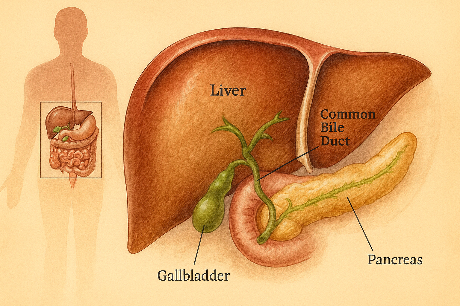
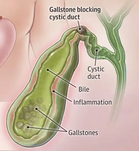
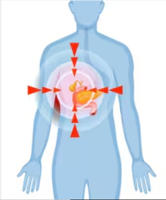
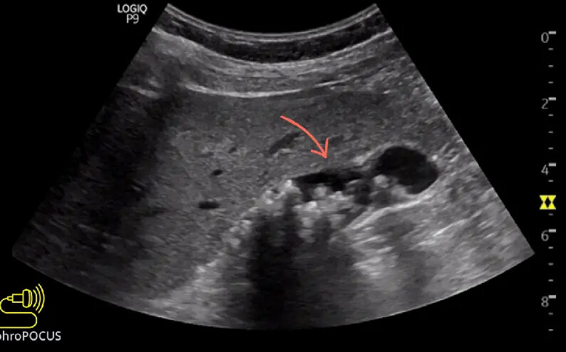
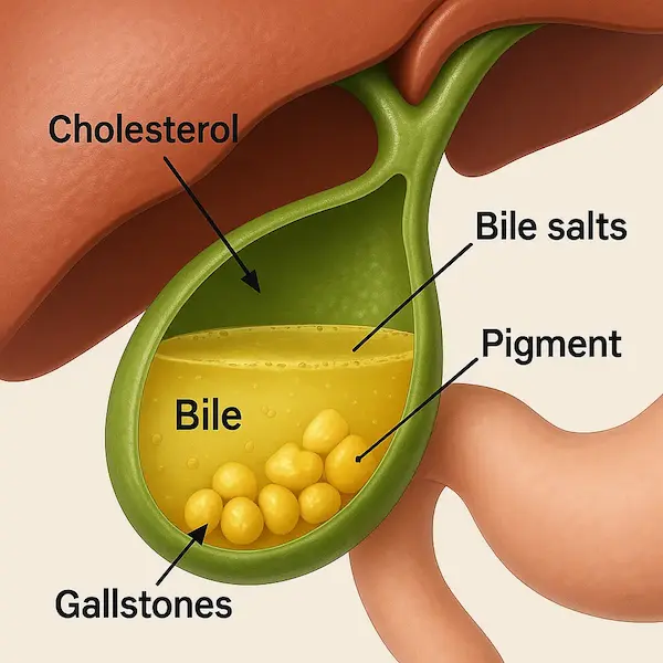
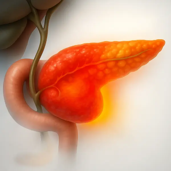
    The gallbladder is a small, pear-shaped organ located just beneath your liver. Its main job is to store bile, a digestive fluid produced by the liver that helps break down fats. Sometimes, the substances in the bile—specifically cholesterol and bilirubin—harden and form pebble-like deposits. These are gallbladder stones.

    They can range in size from a grain of sand to a golf ball. Some people develop just one large stone, while others may have hundreds of tiny ones.

    Why do they form?

    While the exact cause is not always clear, doctors agree that your risk increases if:

     * Your bile contains too much cholesterol or bilirubin.

     * Your gallbladder doesn’t empty correctly or often enough.

     * You consume a diet high in fat and low in fibre.

    Recognizing the Symptoms: When to See a Doctor

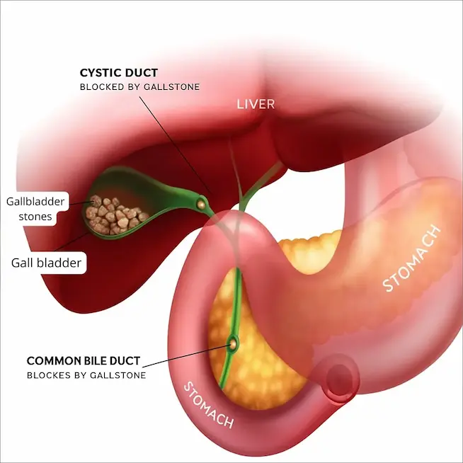
    One of the trickiest aspects of gallstones is that many people have them without knowing it. These are called “silent stones.” However, when a stone blocks a bile duct, the symptoms can be sudden and severe.

    Watch out for these warning signs:

     * Sudden, intensifying pain: Usually in the upper right portion of your abdomen or the centre of your abdomen, just below the breastbone.

     * Referred pain: Pain that radiates to your right shoulder or between your shoulder blades.

     * Nausea and vomiting: Often accompany the pain.

     * Digestive issues: Bloating, indigestion, and intolerance to fatty foods.

    ChatGPT Image Nov 13, 2025, 05 58 43 PM

    If you notice yellowing of your skin or eyes (jaundice) or have a high fever with chills, this constitutes a medical emergency. It suggests the stone has caused an infection or completely blocked the bile duct.

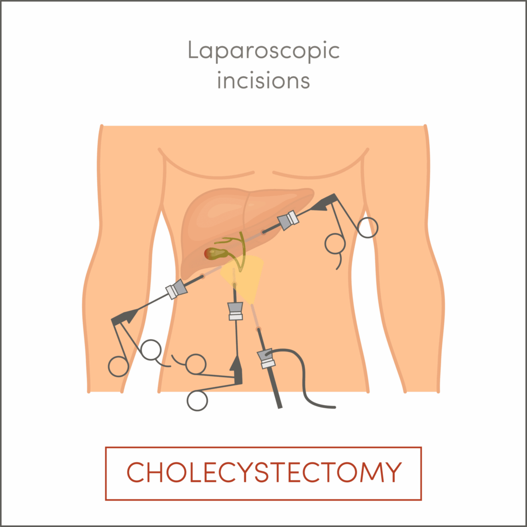
    The Gold Standard Treatment: Laparoscopic Cholecystectomy

    If your stones are causing symptoms, surgery is the only permanent cure. The medical term for gallbladder removal is Cholecystectomy.

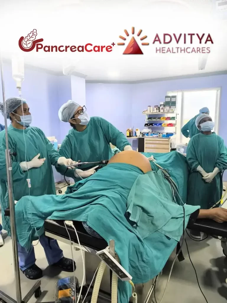
    In the past, this meant open surgery with a large incision and a long recovery. Today, at PancreaCare, we specialise in Laparoscopic Cholecystectomy (Keyhole Surgery).
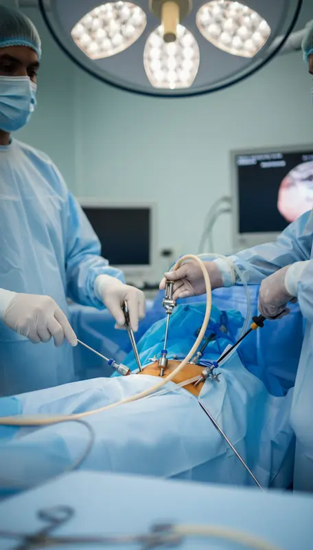
    How it works:

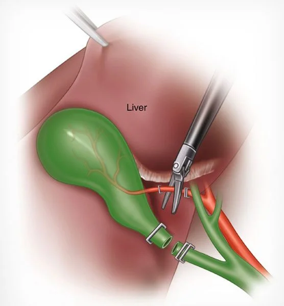
    Instead of a large cut, the surgeon makes 3 to 4 tiny incisions (less than 1 cm) in the abdomen. A laparoscope (a thin tube with a camera) is inserted, allowing the surgeon to see inside your body on a high-definition monitor. The gallbladder is then carefully removed through one of the small incisions.

    IMG 20251114

    Benefits of Laparoscopic Surgery:

     * Minimal Pain: Significantly less post-operative pain compared to open surgery.

     * Quick Recovery: Most patients are discharged within 24 to 48 hours and can return to normal activities within a week.

     * Cosmetic Advantage: The tiny scars fade over time and are barely visible.

     * Safety: It is one of the most commonly performed and safest surgeries in the world when done by experts.

    Busting Common Myths About Gallbladder Surgery

    There is a lot of “advice” floating around in WhatsApp groups and neighbourhood chats in Ranchi and Bokaro. Let’s set the record straight with medical facts.

    Myth 1: “I can flush out gallstones by drinking oil, apple juice, or herbal mixtures.”

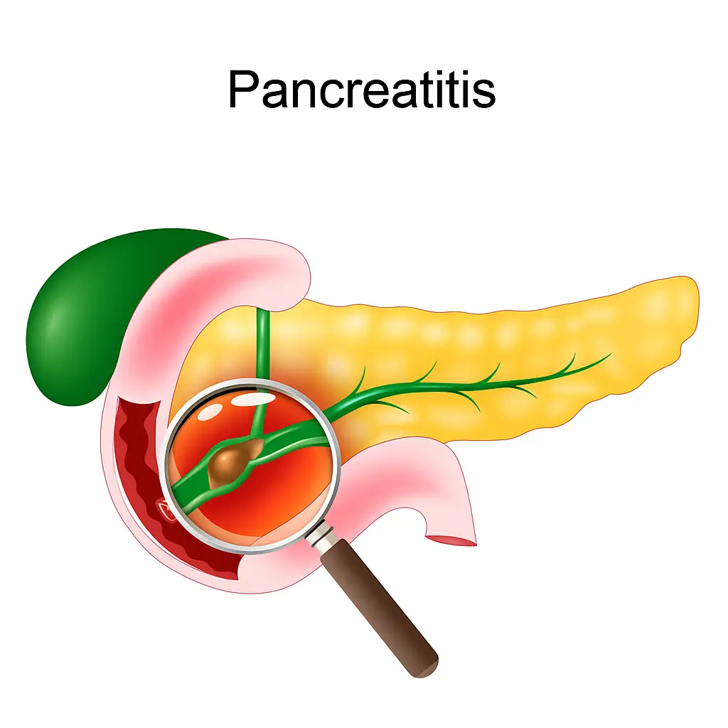
    > Fact: This is a dangerous myth. Gallstones are solid, hard deposits. Drinking oil or juices will not dissolve them. In fact, consuming large amounts of oil to “flush” the system can trigger a severe gallbladder attack or even pancreatitis (inflammation of the pancreas), which is life-threatening.

    Myth 2: “Only the stones need to be removed, not the whole gallbladder.”

    > Fact: Unlike kidney stones, we cannot simply remove gallstones and leave the organ. If we leave the gallbladder, the underlying issue (imbalanced bile) remains, and stones will almost certainly form again quickly. Removing the gallbladder is the only permanent cure.

    Myth 3: “I won’t be able to digest food or eat normal meals after surgery.”

    > Fact: Your liver produces bile, not the gallbladder. The gallbladder just stores it. After surgery, the liver continues to make bile, which drips continuously into your digestive system. You can absolutely eat a normal, healthy diet. You might need to limit very fatty foods for a few weeks while your body adjusts, but you will not have long-term digestive handicaps.

    Myth 4: “Surgery is risky for older patients.”

    > Fact: On the contrary, delaying surgery in older patients can be riskier. An untreated gallbladder can burst or cause severe infection, leading to complex emergency surgeries. Planned (elective) laparoscopic surgery is very safe for elderly patients when performed by specialists like the team at PancreaCare.

    Myth 5: “Laser surgery is different from Laparoscopic surgery.”

    > Fact: Many people in Jharkhand use the term “Laser” operation colloquially for Laparoscopic (Keyhole) surgery. True laser is rarely used for removing the gallbladder itself. The minimally invasive “keyhole” technique is the standard of care you are looking for.

    Why Choose PancreaCare by Advitya Healthcares?

    For years, families in Ranchi, Bokaro, and surrounding districts felt they had to travel to Delhi, Mumbai, or Vellore for complex stomach and liver surgeries. PancreaCare By Advitya Healthcares was born to change that narrative.

    PancreaCare is not just a clinic; it is a centre of excellence dedicated to Gastrointestinal (GI) and Hepato-Pancreato-Biliary (HPB) surgeries.

    Here is why we are the trusted choice in Jharkhand:

     * Specialist Expertise: We are not just general surgeons; we are super-specialists in the digestive system. Our team handles everything from routine gallbladder stones to complex pancreatic cancers with metropolitan-level precision.

     * Advanced Technology: We utilise the latest laparoscopic 4K imaging and surgical equipment, ensuring safer procedures and faster healing for our patients.

     * Local Accessibility: We have brought expert care closer to you. With convenient locations in Ranchi (Morabadi and Bariatu Road) and partnerships in Khunti, you receive premium healthcare without the stress and cost of travelling to a metro city.

     * Patient-Centric Approach: We understand that surgery can be scary. Our team is known for empathetic counselling, explaining every step of the procedure to the patient and their family in simple terms.

     * Affordable Excellence: We believe high-quality healthcare should be accessible. We offer transparent pricing and work with various insurance providers to make your treatment stress-free.

    Untitled design (3)

    Conclusion

    Do not ignore that recurring pain in your abdomen. Living with gallbladder stones is like living with a ticking time bomb that could lead to infection or jaundice at any moment.

    If you reside in Ranchi, Bokaro, Khunti, or anywhere in Jharkhand, expert help is just a phone call away. You do not have to live with pain or rely on unverified home remedies. Trust the specialists who understand your body best.

    Take the Next Step for Your Health:

    Are you or a loved one experiencing symptoms of gallbladder stones? Book a consultation with PancreaCare by Advitya Healthcares today.

     * Visit us in Ranchi, Jharkhand & Bokaro

     * Call us or WhatsApp for an appointment:

    Untitled (200 x 200 px) (50 x 50 px) (2) +91 9211221551

    Untitled (200 x 200 px) (50 x 50 px) (2) +91 9211221552

    Untitled (200 x 200 px) (50 x 50 px) (2) +91 9211221553

    Untitled (200 x 200 px) (50 x 50 px) (2) +91 9211221554

     * Website:   https://advityahealthcares.com/

    PancreaCare by Advitya Healthcares – bringing world-class surgical care home to Jharkhand.

  • Expert Advice for Complete Recovery After Pancreatic Cancer – From PancreaCare By Advitya Healthcares

    Expert Advice for Complete Recovery After Pancreatic Cancer – From PancreaCare By Advitya Healthcares

    By PancreaCare By Advitya Healthcares — complete solutions for all pancreas problems in Ranchi, Jharkhand, Bokaro

    If you or a loved one in Ranchi, Jharkhand, Bokaro is aiming for a full recovery after pancreatic cancer, a clear roadmap makes every decision easier. This guide explains the typical sequence—from diagnosis and surgery to nutrition, adjuvant therapy, rehab, and follow-up—so you know what to expect and how PancreaCare By Advitya Healthcares supports you locally in Ranchi, Jharkhand, Bokaro and nearby areas (Harmu, Doranda, Bariatu, Morabadi, Kanke, Namkum, Ratu Road, Hinoo).

    1) Staging and first visit in Ranchi, Jharkhand, Bokaro

    Recovery starts with accurate staging. At your first visit in Ranchi, Jharkhand, Bokaro, we review prior reports and arrange protocol-driven imaging: contrast CT, MRI/MRCP, and EUS when needed. Blood tests (including CA 19-9) help classify disease as resectable, borderline resectable, locally advanced, or metastatic. A multidisciplinary tumor board (surgery, medical oncology, radiation oncology, radiology, gastroenterology, pathology, dietetics) then decides the safest, most effective plan—surgery first, neoadjuvant therapy, or a clinical trial. Getting this step right avoids delays and unnecessary travel outside Ranchi, Jharkhand, Bokaro.

    2) Neoadjuvant therapy—when treatment comes before surgery

    Neoadjuvant Theraphy Image by PancreaCare By Advitya Healthcares
    Neoadjuvant Theraphy Image by PancreaCare

    If the tumor touches key blood vessels or the biology looks aggressive, chemotherapy (sometimes chemoradiation) before surgery can shrink disease, treat micrometastases early, and test responsiveness. We track progress with scans every 8–12 weeks and trend CA 19-9. Many Ranchi, Jharkhand, Bokaro patients appreciate that this phase can be coordinated close to home, with our team guiding side-effect control, enzyme timing, and nutrition.

    3) Curative surgery—your best chance at long-term control

    For operable cases, surgery offers the strongest chance of durable remission. The operation depends on location:

    • Whipple (pancreaticoduodenectomy) for head/uncinate tumors
    • Distal pancreatectomy (often with splenectomy) for body/tail tumors
    • Total pancreatectomy in selected situations

    What recovery looks like: monitored post-op care, early walking, breathing exercises, and a stepwise return to oral diet. Enhanced-Recovery-After-Surgery (ERAS) pathways—pain control, anti-clot measures, early feeding when safe—help reduce complications and speed discharge. If you live outside central Ranchi, Jharkhand, Bokaro, we coordinate logistics so follow-ups remain smooth.

    Nutrition, enzymes, and glucose after surgery

    Because surgery changes how bile and enzymes mix with food, some people develop pancreatic exocrine insufficiency (PEI)—oily stools, gas, bloating, and weight loss despite good intake. Pancreatic enzyme replacement therapy (PERT), taken with meals/snacks, restores digestion, improves energy, and supports weight gain. If a large portion of the pancreas is removed, blood sugar can fluctuate; an individualized diabetes plan (monitoring, diet, tablets/insulin) keeps it steady. Our Ranchi, Jharkhand, Bokaro dietitians design small, frequent, protein-rich meals, hydration goals, and vitamin support tailored to your tolerance.

    4) Adjuvant therapy—lowering the risk of recurrence

    After sufficient healing (commonly 6–8 weeks post-op), most patients are considered for adjuvant chemotherapy to reduce relapse risk. Regimens, duration, and start dates depend on stage, margins, nodes, and overall fitness. If you already received neoadjuvant therapy, your post-op plan is adjusted to complete the optimal total course. Our Ranchi, Jharkhand, Bokaro team focuses on timely starts, proactive side-effect management, and maintaining strength so you can finish treatment as planned.

    5) Follow-up & surveillance

    A structured follow-up plan is your safety net:

    Pancreatic Cancer Follow-up and Surveiliance
    Pancreatic Cancer Follow-up and Surveiliance
    • Every 3–6 months for years 0–2: clinic review, labs, CA 19-9, and CT/MRI as indicated
    • Every 6–12 months for years 3–5 (or as advised)
      Between visits, call if you notice new jaundice, persistent back/abdominal pain, unexplained weight loss, greasy stools, or new-onset diabetes. We also provide survivorship guidance—vaccines if your spleen was removed, bone/muscle health, return-to-work planning, and mental-health support right here in Ranchi, Jharkhand, Bokaro.

    6) Day-to-day recovery: rehab, lifestyle, and support

    • Activity: Begin with short walks and breathing exercises; build to light resistance and aerobic sessions as cleared. Movement improves gut motility, sleep, and mood.
    • Diet: Protein-forward meals; trial fats carefully; time expandwith food. Consider lactose-free or low-fat options if symptomatic.
    • Tracking: Keep a simple diary (stools, weight, appetite, glucose). It helps your clinicians fine-tune enzymes, nutrition, and medicines.
    • Emotional health: Fatigue and “scanxiety” are common. Counseling, caregiver involvement, and peer groups in Ranchi, Jharkhand, Bokaro can make a real difference.


    Why choose PancreaCare By Advitya Healthcares in Ranchi, Jharkhand, Bokaro

    • End-to-end pancreas care in Ranchi, Jharkhand, Bokaro: from first consult to survivorship
    • Evidence-based diagnostics: CT/MRI/MRCP/EUS with standardized protocols
    • Advanced endoscopy & stenting for jaundice/obstruction
    • High-volume pancreatic surgery with ERAS pathways
    • Medical & radiation oncology tailored to your case
    • Nutrition, pain & diabetes care, and structured follow-up
    Doctors Of PancreaCare By Healthcares
    Doctors at PancreaCare By Healthcares

    If you’re in Ranchi, Jharkhand, Bokaro and experiencing persistent upper abdominal/back pain, jaundice, greasy stools, weight loss, or new-onset diabetes, don’t delay—early evaluation changes outcomes.


    Call to action (Ranchi, Jharkhand, Bokaro)

    Book an in-person or video consult with PancreaCare By Advitya Healthcares in Ranchi, Jharkhand, Bokaro. We’ll map your sequence—diagnostics → treatment → rehab → surveillance—and walk with you through every milestone.


  • Liver Cancer Awareness Month: Understanding Risk, Detection, and Care

    Liver Cancer Awareness Month: Understanding Risk, Detection, and Care

    October highlights Liver Cancer Awareness Month, an important opportunity to raise awareness of one of the fastest-growing cancer diagnoses worldwide. As liver cancer cases increase in many areas, early detection, prevention, and informed treatment decisions are crucial for improving patient outcomes.

    This article provides a comprehensive overview of risk factors, causes, staging, clinical symptoms, diagnostic tools, treatment options, and links to aftercare resources for individuals and families navigating liver cancer.

    Etiology and Risk Factors

    A green awareness ribbon Pancreas & Advitya Healthcares Ranchi and kolkata

    Liver cancer primarily develops in individuals with underlying liver disease. The most common type, hepatocellular carcinoma (HCC), arises from hepatocytes (the primary liver cells). Intrahepatic cholangiocarcinoma, a cancer of the bile ducts within the liver, is less common but clinically significant.

    Key risk factors include:

    • Chronic viral hepatitis (HBV, HCV)
    • Cirrhosis (from alcohol, viral hepatitis, or non-alcoholic steatohepatitis)
    • Heavy alcohol consumption
    • Non-alcoholic fatty liver disease (NAFLD)
    • Obesity and Type 2 diabetes
    • Tobacco use
    • Exposure to aflatoxins (naturally occurring toxins in certain foods)
    • Family history of liver cancer
    • Hemochromatosis and other inherited liver disorders

    Preventive strategies such as hepatitis B vaccination, harm reduction for hepatitis C transmission, and lifestyle modification play a critical role in reducing liver cancer risk.

    Pathogenesis and Causes

    Liver cancer typically develops in the setting of chronic inflammation and hepatocellular injury, which promotes DNA damage, fibrosis, and eventual malignant transformation of liver cells.

    Key pathological processes include:

    • Chronic hepatitis (viral or autoimmune)
    • Fibrosis and cirrhosis progression
    • Cellular dysplasia within regenerative nodules
    • Genetic mutations or epigenetic alterations

    Understanding these mechanisms helps develop targeted therapies and surveillance protocols for high-risk populations.

    Cancer Staging

    Classification of hcc and its characteristics based on the bclc staging system hcc can Pancreacare logo with white background

    Accurate staging guides treatment and prognostication. Most institutions use the Barcelona Clinic Liver Cancer (BCLC) staging system, which considers tumour burden, liver function, performance status, and cancer-related symptoms.

    General Stages:

    • Stage 0 (Very Early): Single small tumour (<2 cm), preserved liver function
    • Stage A (Early): Single or up to three nodules <3 cm, no vascular invasion
    • Stage B (Intermediate): Multiple tumours without vascular invasion
    • Stage C (Advanced): Portal vein invasion or extrahepatic spread
    • Stage D (End-Stage): Severely impaired liver function, poor performance status

    Staging also considers the Child-Pugh score and the Model for End-Stage Liver Disease (MELD) in therapeutic decision-making.

    Signs and Clinical Presentation

    Clinical representation of a patient experiencing liver cancer symptoms, including abdominal pain and jaundice, with anatomical highlights Advitya healthcares

    Early-stage liver cancer may be asymptomatic. When symptoms do emerge, they often indicate disease progression:

    • Right upper quadrant abdominal pain or fullness
    • Unexplained weight loss
    • Anorexia and early satiety
    • Fatigue and weakness
    • Ascites
    • Jaundice (yellowing of skin and sclera)
    • Pruritus
    • Pale stools and dark urine
    • Hepatomegaly or palpable mass

    Due to nonspecific symptoms, high-risk individuals should undergo routine surveillance imaging and blood work

    Diagnostic Approaches

    Clinical representation of a patient experiencing liver cancer symptoms, including abdominal pain and jaundice, with anatomical highlights Advitya healthcares 2

    Diagnosis of liver cancer involves a combination of imaging, laboratory testing, and in some cases, histologic confirmation:

    • Imaging: Multiphasic contrast-enhanced MRI or CT scan to assess arterial enhancement and washout pattern
    • Serum biomarkers: Elevated alpha-fetoprotein (AFP) levels, though not definitive alone
    • Liver biopsy: Generally reserved for indeterminate imaging findings or clinical trials
    • Liver function tests: AST, ALT, ALP, bilirubin, INR to evaluate hepatic reserve

    High-risk patients (e.g., those with cirrhosis or HBV/HCV) should undergo ultrasound screening every 6 months.

    Treatment Modalities

    Icons or infographic showing various treatment options for liver cancer, including surgery, immunotherapy, embolisation, and ablation

    Treatment is individualised based on cancer stage, liver function, patient health, and institutional resources. Options include:

    1. Curative Therapies – add images – add images for right hepatectomy, left hepatectomy, trisegmentectomy

    • Surgical resection: Preferred for localised tumours and adequate liver reserve
    • Liver transplantation: Ideal for patients within the Milan criteria
    • Local ablation: Radiofrequency or microwave ablation for small lesions

    2. Locoregional Therapies

    • Transarterial chemoembolization (TACE)
    • Transarterial radioembolization (TARE)

    3. Systemic Therapies

    • Targeted therapies: Sorafenib, Lenvatinib, Regorafenib, Cabozantinib
    • Immunotherapy: Atezolizumab plus Bevacizumab is a first-line standard
    • Chemotherapy: Limited role; used in select cases

    4. Palliative and Supportive Care

    • Symptom management, nutrition, psychological support

    Multidisciplinary care is essential—often involving hepatologists, oncologists, interventional radiologists, and surgeons

    Survivorship & Aftercare

    Icons or infographic showing various treatment options for liver cancer, including surgery, immunotherapy, embolisation, and ablation

    Long-term follow-up is essential for:

    • Detecting recurrence (imaging, AFP monitoring)
    • Managing comorbid liver disease
    • Supporting physical and emotional recovery
    • Providing nutritional guidance
    • Monitoring for treatment-related complications

    👉 For comprehensive information, visit our previous post:
    Aftercare for Liver Cancer Survivors: A Guide to Ongoing Health and Support

    Conclusion: Awareness Leads to Action

    Icons or infographic showing various treatment options for liver cancer, including surgery, immunotherapy, embolisation, and ablation

    Liver Cancer Awareness Month reminds us of the urgent need for education, screening, and access to care. Increased awareness leads to earlier diagnosis, improved survival, and better quality of life for those affected.

    If you or someone you know is at risk, speak with a healthcare provider about screening options.


    📚 Additional Resources


    Disclaimer: The information provided in this article is for educational purposes only and should not be interpreted as medical advice. Always consult with a qualified healthcare provider for any health concerns or decisions regarding diagnosis and treatment

  • Acute Pancreatitis: A Sudden Inflammation You Can’t Ignore

    Acute Pancreatitis: A Sudden Inflammation You Can’t Ignore

    What is Acute Pancreatitis?

    Acute pancreatitis is a sudden inflammation of the pancreas that develops within hours to days. It ranges from mild, short-lived discomfort to severe, life-threatening illness.

    The pancreas normally releases digestive enzymes into the small intestine. In acute pancreatitis, these enzymes become trapped inside and begin digesting the pancreas itself — causing swelling, pain, and sometimes organ failure.

    Causes of Acute Pancreatitis

    The two most common triggers are:

    • Gallstones → Block the duct and trap enzymes inside.

    • Alcohol use → Heavy drinking irritates and inflames pancreatic tissue.

    Other causes include:

    • High triglycerides (>1000 mg/dL)

    • Certain medications

    • Trauma or post-surgical complications

    • Viral infections (mumps, hepatitis)

    • Genetic predispositions

    Patient eating healthy with enzymes beside plate, caption 'Living Well with Chronic Pancreatitis' Advitya healthcare

    Symptoms

    • Sudden, severe upper abdominal pain (often radiates to the back)

    • Pain worsens after eating, especially fatty meals

    • Nausea and repeated vomiting

    • Fever, swollen belly, rapid pulse

    • Severe cases: difficulty breathing, confusion, low blood pressure

    Patient holding abdomen with pain radiating to back advitya healthcare

    Diagnosis

    • Blood tests: Elevated amylase & lipase

    • Ultrasound: Looks for gallstones, swelling

    • CT/MRI scans: Detects severity and complications

    • ERCP/MRCP: Detailed duct imaging

    Collage of lab reports, ultrasound, CT scan Advitya healthcare

    Treatment & Hospital Care

    Most patients need hospitalization. Treatment includes:

    • IV fluids

    • Pain relief

    • Bowel rest (no food initially)

    • Gallstone removal if needed

    • Antibiotics or drainage for infection

    • ICU care for severe cases

    : ICU care illustration

    Complications

    • Pancreatic necrosis

    • Abscesses or pseudocysts

    • Organ failure (lungs, kidneys, heart)

    • Sepsis

    : ICU care illustration

    Takeaway

    Acute pancreatitis is a medical emergency. If you have sudden, severe abdominal pain, seek care immediately. Early treatment saves lives.

    Doctor reassuring patient in ER with caption 'Act Early. Save Your Pancreas Advitya Healthcrae
  • Fatty Pancreas: The Silent Condition You Shouldn’t Ignore

    Fatty Pancreas: The Silent Condition You Shouldn’t Ignore


    Most people have heard of fatty liver disease, but did you know fat can also build up inside your pancreas?
    This is called fatty pancreas or pancreatic steatosis. For many, it causes no obvious symptoms at first — which is why it’s often called a silent condition. But ignoring it can lead to serious health risks, including diabetes, pancreatitis, and even pancreatic cancer in long-standing cases.

    What is a Fatty Pancreas?

    Your pancreas sits deep in the abdomen and has two main jobs:

    • Producing digestive enzymes that help break down food

    • Releasing hormones like insulin that control blood sugar

    When fat accumulates inside this organ, it can interfere with both. Doctors often find fatty pancreas together with fatty liver, since both conditions share common risk factors.

    Advitya healthcare normal vs fatty pancreas

    Why Does Fat Build Up in the Pancreas?

    Fatty pancreas is usually the result of metabolic and lifestyle factors rather than a single cause. Risk factors include:

    • Obesity or weight gain around the belly

    • Diets high in fried, processed, or sugary foods

    • Alcohol overuse

    • Lack of exercise

    • Type 2 diabetes and insulin resistance

    • Fatty liver disease (they often coexist)

    • Aging and slowed metabolism

    Advitya healthcare lifestyle

    Who is at Risk?

    You may have a higher chance of developing fatty pancreas if you:

    • Struggle with weight gain or central obesity

    • Have type 2 diabetes

    • Have high cholesterol or triglycerides

    • Drink alcohol regularly

    • Have high blood pressure

    • Have a family history of metabolic diseases

    Advitya healthcare Collage

    Symptoms – Why It’s Silent

    In the early stages, fatty pancreas is usually asymptomatic. Most cases are discovered by accident on an ultrasound or CT scan.

    When symptoms do occur, they may include:

    • Dull upper abdominal discomfort

    • Nausea, bloating, indigestion

    • Sudden blood sugar fluctuations

    • Fatigue or weakness

    • Episodes of pancreatitis (in some cases)

    Advitya healthcare Symptom slider

    How is it Diagnosed?

    • Ultrasound → First-line, often shows fatty changes

    • CT or MRI → Detailed view of pancreatic tissue

    • MRCP → Evaluates pancreatic and bile ducts

    • Blood tests → Check sugar, cholesterol, triglycerides, enzymes

    Advitya healthcare

    Can it be Treated?

    There’s no single pill for fatty pancreas. But it can often be stabilized — and sometimes reversed — with lifestyle changes:

    • Lose weight gradually with balanced nutrition

    • Eat more fruits, vegetables, whole grains, and lean proteins

    • Avoid fried, processed, and sugary foods

    • Exercise regularly (30–40 minutes most days)

    • Limit or avoid alcohol

    • Control diabetes, cholesterol, and blood pressure

    • Regular check-ups with a gastroenterologist

    Advitya healthcare Lifestyle

    Why It Matters – The Risks of Ignoring It

    A fatty pancreas may look harmless, but evidence shows it can increase the risk of:

    • Type 2 diabetes – studies link pancreatic fat to reduced insulin secretion

    • Pancreatitis – fat deposits may worsen inflammation

    • Pancreatic cancer – long-standing fatty pancreas may raise cancer risk

    Advitya healthcare Lifestyle 1

    Key Takeaway

    Fatty pancreas is silent but not harmless.
    With early detection, healthy habits, and medical monitoring, you can protect your pancreas and lower your risk of serious complications.

    Advitya healthcare Lifestyle 2
  • Pancreatic Cancer: The Silent Killer

    Pancreatic Cancer: The Silent Killer

    Pancreatic cancer is one of the most feared cancers of the digestive system. Often called the “silent killer”, it develops quietly with almost no early symptoms. By the time it’s discovered, the disease is usually advanced. That’s why awareness, timely check-ups, and early detection are so critical.

    Gemini generated image ay2kobay2kobay2k

    What is Pancreatic Cancer?

    Pancreatic cancer begins when abnormal cells in the pancreas grow uncontrollably. The pancreas, hidden deep in the abdomen, has two main jobs:

    • Producing digestive enzymes to help break down food

    • Producing hormones like insulin to regulate blood sugar

    The most common type is pancreatic adenocarcinoma, arising from the ductal cells of the pancreas.

    Whatsapp image 2025 10 05 at 6.26.25 pm

    Causes and Risk Factors

    There’s no single cause, but certain factors raise the risk:

    • Age: Most cases occur after 60

    • Smoking: Strongest preventable risk factor

    • Chronic pancreatitis: Long-term inflammation of the pancreas

    • Diabetes: Especially new-onset after age 50

    • Obesity and sedentary lifestyle

    • Family history/genetics: BRCA2, Lynch syndrome

    • Alcohol excess: Via chronic pancreatitis and liver injury

    • Diet: High in processed/red meats, low in fruits/vegetables

    Dark blue technology sales percentage graph

    Symptoms: Why Early Detection is Hard

    • Persistent upper abdominal or back pain

    • Unexplained weight loss

    • Loss of appetite, bloating, indigestion

    • Jaundice: yellow skin/eyes, dark urine, pale stools

    • New-onset diabetes in adults >50 years (not obese)

    • Fatigue and weakness

    Diagnosis

    • Imaging: CT, MRI, PET scans

    • Endoscopic Ultrasound (EUS): Precise imaging, biopsy possible

    • ERCP: Examines ducts, allows stenting and tissue sampling

    • Blood tests: CA 19-9 tumor marker (not always specific)

    • Biopsy: Final confirmation under microscope

    Cream colorful abstract flowchart infographic chart

    Treatment Options

    Treatment depends on stage and patient health:

    1) Surgery

       • Whipple procedure (head of pancreas)

       • Distal pancreatectomy (tail/body)

       • Total pancreatectomy (rare, extensive disease)

    2) Chemotherapy – neoadjuvant (before surgery), adjuvant (after surgery), or for advanced disease

    3) Radiation therapy – often combined with chemotherapy

    4) Targeted therapy & Immunotherapy – for specific mutations and selected cases

    5) Palliative care – pain relief and symptom management for advanced stages

    Cream colorful abstract flowchart infographic chart (1)

    Prognosis & Why Early Detection Matters

    While pancreatic cancer is serious, outcomes are significantly better when detected early and treated fully:

    • Patients who undergo curative surgery plus chemotherapy achieve 5-year survival rates of 20–30%, sometimes even up to 35–40% in specialized centers.

    • Unfortunately, many patients are diagnosed at advanced stages, when curative surgery isn’t possible. This lowers the overall average survival to around 10–12%.

    Whatsapp image 2025 10 05 at 7.22.33 pm

    Key Takeaways

    • Pancreatic cancer is often silent early on but dangerous if ignored.
    • Risk factors include smoking, obesity, diabetes, and family history.
    • Warning signs: jaundice, unexplained weight loss, and sudden diabetes.
    • Early detection saves lives — consult a gastroenterologist if you have persistent risk factors.

    Gemini generated image no41yrno41yrno41
  • Gallbladder Stones

    Gallbladder Stones

    What Is the Gallbladder?

    Think of your liver as a factory. This factory makes a special liquid called bile which helps digest (break down) fats in your food.

    Now, this bile needs a storage place. That storage place is the gallbladder — a small pouch sitting under the liver.

    📦 The gallbladder is just like a storage tank. It doesn’t make bile, it only stores it. When you eat food, the gallbladder squeezes and sends bile into the intestine to help digest fats.

    What Are Gallbladder Stones?

    Sometimes, the bile stored inside the gallbladder becomes too thick or too rich in cholesterol. Slowly, it starts forming hard lumps — these are called gallstones.

    These stones can be:
    As small as sand
    Or as big as a marble

    Some people never know they have stones. Others may get pain and health problems because of them.


    Why Do Gallstones Form?

    Gallstones form when there is:

    Imagine keeping milk in a pot without stirring. After some time, a cream layer forms. Similarly, bile sitting for too long in the gallbladder can form stones.


    What Problems Can Gallstones Cause?

    7e269190 94df 4471 8c15 0e0b9cb91461
    • Bile duct blockage (Choledocholithiasis): Severe pain with jaundice and dark urine.
    Image 1
    • Pancreatitis: If a stone blocks the pancreatic duct, it can cause dangerous inflammation of the pancreas.
    Chatgpt image sep 6, 2025, 06 35 45 pm
    • Gallstone attack & rare severe complications: Sudden, intense pain (often after fatty/oily meals). In very rare cases, stones can lead to life-threatening issues like gallbladder ruptur
    Image

    Not everyone with gallstones has symptoms. Many people live with them unknowingly.
    But when gallstones block the flow of bile, they can cause problems.
    Most gallstones are harmless, but when they cause symptoms, medical attention is necessary.

    Common symptoms

    This pain is sometimes called a gallbladder attack.


    Who Gets Gallstones More Often?


    How Do Doctors Find Gallstones?

    9c0bb605 d070 434b 941f 90f4e87a7c6d

    Doctors usually do an ultrasound (a safe, simple test) to see stones.

    Other tests may include:


    How Is Surgery Done?

    Cholecystectomy laparascopic incisions

    Why the Gallbladder Is Removed Instead of Just the Stones?

    Gallbladder surgery image

    Can You Live Without a Gallbladder?

    The answer is YES !!!

    The liver, not the gallbladder, is responsible for producing bile. The gallbladder merely serves as a storage reservoir. After surgery, bile continues to flow directly from the liver into the intestine through the bile duct, ensuring that digestion is not interrupted.
    In practical terms, the liver functions as the factory, the gallbladder as the storage tank, and the bile duct as the pipeline. Even if the storage tank is removed, the pipeline remains intact and bile continues to reach the intestine.
    Most patients recover within a few weeks and return to normal digestion. A small proportion, around 5–10%, may experience temporary symptoms such as bloating, gas, or mild loose motions, which typically resolve with time.

    How to Prevent Gallstones?

    Chatgpt image sep 6, 2025, 06 49 55 pm

    You can reduce the risk by:

    – Eating a balanced diet with less oil and more fruits, vegetables, and fiber
    – Avoiding crash diets or sudden weight loss
    – Maintaining a healthy weight
    – Exercising regularly
    – Controlling diabetes and cholesterol

    Key Takeaways

    Gallstones are common, especially in India.

    Not all gallstones need treatment — only when they cause problems.

    If surgery is needed, removing the gallbladder is safe and the body adapts well.

    You can live a completely normal, healthy life without a gallbladder.

  • Gallbladder Cancer

    Gallbladder Cancer

    Gallbladder cancer is a rare but serious disease that begins in the gallbladder — a small, pear-shaped organ located beneath your liver. The gallbladder’s main job is to store bile, a fluid that helps digest fats. Because the gallbladder is tucked away deep inside the body, cancer here can often go unnoticed until it is more advanced.


    Why Early Detection Is Challenging

    Chatgpt image jul 25, 2025, 12 38 33 pm

    Gallbladder cancer often doesn’t cause clear symptoms in its early stages. When symptoms do appear, they can be mistaken for other digestive issues, such as gallstones or infections. This can delay diagnosis and treatment.

    At the cellular level, cancer develops when normal cells lose their control signals, multiply uncontrollably, and form a lump or mass. These abnormal cells can invade nearby tissues and spread to other organs — a process called metastasis.


    Common Symptoms

    While having these symptoms does not always mean cancer, it’s important to discuss them with your healthcare provider if they persist:

    • Persistent abdominal pain in the right upper quadrant (often dull)
    • Jaundice (yellowing of the skin or eyes)
    • Nausea or vomiting
    • Loss of appetite and unexplained weight loss
    • Fever or general fatigue

    In some cases, symptoms occur when the tumor presses on the bile duct or liver.


    What has been found to be associated with gall bladder cancer?

    Certain factors may increase the risk of gallbladder cancer, including:

    • Gallstones — especially large stones present for many years that can damage the gallbladder wall
    • Chronic gallbladder inflammation
    • Typhoid can persist in asymptomatic long-term carriers.
    • Being female (women are affected more often than men in India)
    • Older age (most cases occur in people over 65)
    • Family history of gallbladder disease
    • Certain ethnic or geographic populations (Native American, Hispanic, and some Indian regions)

    How It’s Diagnosed

    If gallbladder cancer is suspected, your doctor may recommend:

    • Ultrasound — first-line imaging to detect abnormalities
    • CT scan or MRI — to assess wall thickening, unhealthy appearance, or lumps, and to check if nearby lymph nodes or the liver are involved
    • Blood tests — to evaluate liver function
    • Biopsy — to confirm the presence of cancer cells

    No imaging test (CT, MRI, PET) is 100% accurate in differentiating between cancerous and non-cancerous thickening. This is why, in some instances, surgical removal of the gallbladder is advised for confirmation.

    If facilities are available, the removed gallbladder can be examined during surgery via a frozen section test. If cancer is confirmed, the surgeon may remove part of the liver and surrounding lymph nodes as part of definitive treatment.


    Treatment Options

    Treatment depends on the stage of the disease and overall health:

    • Surgery — Primary treatment for early-stage disease. This may include removal of the gallbladder, part of the liver, and nearby lymph nodes. For gallbladder cancer that has not spread beyond the gallbladder and nearby tissues, surgery offers the best chance for cure. The standard procedure for most patients with resectable disease beyond very early stage is a radical cholecystectomy.

    What Does a Radical Cholecystectomy Include?

    Ec use4xgaa3tki
    1. Removal of the gallbladder
    2. Liver resection — The surgeon removes part of the liver where the gallbladder is attached, usually:
      • Segments IVb and V (about 2–3 cm depth of liver tissue)
      • This ensures removal of any microscopic cancer spread into the liver bed.
    3. Lymph node removal (lymphadenectomy)
      • Nodes in the hepatoduodenal ligament (around the bile duct, hepatic artery, and portal vein) are removed.
      • Guidelines recommend at least 6 lymph nodes be retrieved for accurate staging.
    4. Bile duct removal
      • Not routinely performed.
      • Done only if cancer directly invades the bile duct or is too close to achieve a safe margin without removal.
    5. Chemotherapy —. Chemotherapy uses medicines to kill cancer cells or stop them from growing. In gallbladder cancer, it is used in different settings:

    Adjuvant Chemotherapy (After Surgery) – Given for 3–6 months after radical cholecystectomy (stage II or higher, or with high-risk features) to kill hidden cancer cells and lower recurrence risk. Standard is capecitabine for 6 months (BILCAP trial); gemcitabine + cisplatin may be used if not suitable.

    Neoadjuvant Chemotherapy (Before Surgery) – Used in selected borderline or locally advanced cases to shrink the tumor and make surgery possible. Common regimens include gemcitabine + cisplatin.

    Sometimes, gallbladder cancer is discovered unexpectedly after surgery for gallstones. If it’s detected early, no further treatment may be needed. If more advanced, a second surgery may be recommended.


    Incidental Gallbladder Cancer (IGBC): What You Should Know

    What is Incidental Gallbladder Cancer?

    Incidental Gallbladder Cancer (IGBC) is gallbladder cancer that is discovered unexpectedly—usually after a gallbladder removal surgery (cholecystectomy) that was performed for another reason, such as gallstones or inflammation (cholecystitis).
    The diagnosis is often made when the removed gallbladder is examined under a microscope by a pathologist.


    Why does it happen?

    Gallbladder cancer is uncommon, and in its early stages, it rarely causes symptoms different from gallstones.
    Because of this, cancer may go unnoticed until surgery is done for what seems like a benign problem.
    Risk factors include:

    • Long-standing gallstones
    • Gallbladder polyps (especially >1 cm)
    • Chronic gallbladder inflammation (porcelain gallbladder)
    • Certain genetic and environmental factors

    What happens after the diagnosis?

    If IGBC is detected, your doctor will review:

    • Stage of cancer (how deep it has invaded the gallbladder wall and whether it has spread)
    • Margins (whether cancer cells are present at the edge of the removed tissue)
    • Lymph node status (if available)

    Further treatment may include:

    • Additional surgery (radical cholecystectomy with liver wedge resection and lymph node removal) – often advised if the cancer is stage T1b or higher.
    • Imaging scans (CT/MRI) to check for spread.
    • Oncology referral for chemotherapy if the disease is advanced or surgery is not possible.

    Why is timely action important?

    Gallbladder cancer can spread quickly to the liver and surrounding areas.
    If IGBC is detected early and treated appropriately, the chances of long-term survival improve significantly.
    Delaying evaluation or treatment can allow the disease to progress, limiting treatment options.


    Key Takeaways for Patients

    • IGBC is often found unexpectedly after gallbladder surgery.
    • Early-stage IGBC can often be treated successfully with timely surgery.
    • Always review your gallbladder histopathology report after surgery.
    • If IGBC is reported, consult a hepatobiliary or gastrointestinal cancer surgeon promptly.

    Living With and Beyond Gallbladder Cancer

    A gallbladder cancer diagnosis can feel overwhelming, but advances in surgical techniques, chemotherapy, and supportive care have improved outcomes. Early detection, timely surgery, and close follow-up care remain key to improving survival and quality of life.

  • Understanding Abdominal Sarcomas: What a patient should know?

    Understanding Abdominal Sarcomas: What a patient should know?

    What Are Abdominal Sarcomas?

    Advitya healthcares understanding abdominal sarcomas

    Abdominal sarcomas are rare cancers that originate from the connective tissues within the abdomen, such as fat, muscle, nerves, fibrous tissue, or blood vessels. Unlike the more common cancers that start in organs like the colon or pancreas, sarcomas develop in the soft tissues or mesenchymal tissue, which forms structural and supportive parts of the body.

    These tumours are classified into two major groups:

    • Retroperitoneal Sarcomas: These develop in the space behind the abdominal organs, known as the retroperitoneum.
    • Intra-abdominal Sarcomas: Arise within abdominal organs or the abdominal wall.

    Though sarcomas are rare (making up <1% of all adult cancers), they can behave aggressively and require specialised care.

    What are the causes of Abdominal Sarcomas?

    • Genetic Conditions
      Some people inherit conditions that make them more likely to develop sarcomas. These include genetic syndromes like Li-Fraumeni syndrome and Neurofibromatosis type 1.
    • Radiation Exposure
      People who have received radiation therapy in the past, especially to the abdominal area, may have a higher risk of developing soft tissue sarcomas years later.
    • Chemical Exposure
      Long-term exposure to certain chemicals such as vinyl chloride, dioxins, or herbicides may increase the risk in some individuals.
    • Chronic Swelling or Injury
      Although rare, long-standing swelling or injury in a certain part of the body may, over time, contribute to the development of sarcomas.
    • Unknown Causes
      In many cases, there is no clear cause. Sarcomas can occur without any family history, exposure, or warning signs.

      Abdominal sarcomas are not caused by lifestyle factors like diet or stress. They are uncommon and can affect anyone. 

    What are the common types of soft tissue sarcomas?

    • Liposarcoma
       This type originates in fat cells and is one of the most common types of abdominal sarcomas. It usually grows slowly but can become large before causing symptoms.
    • Leiomyosarcoma
       This type of cancer begins in smooth muscle tissue, often originating from the walls of the intestines or blood vessels in the abdomen. It may grow more aggressively than some other types.
    • Gastrointestinal Stromal Tumour (GIST)
       GISTs arise from special cells in the wall of the digestive tract. They are a unique type of sarcoma and are often treated differently, sometimes with targeted medicines.
    • Undifferentiated Pleomorphic Sarcoma (UPS)
       Previously called malignant fibrous histiocytoma, this is a high-grade tumour that doesn’t resemble any specific tissue type. It can occur in the retroperitoneum (deep part of the abdomen).
    • Desmoid Tumours (Aggressive Fibromatosis)
       Although not considered a true sarcoma or cancer, desmoid tumours arise from connective tissue and can grow aggressively. They do not spread to other organs, but they can invade nearby tissues and sometimes recur after treatment. Management often includes surgery, medications, or close monitoring depending on the case.
    • Other Rare Types
       These include synovial sarcoma, angiosarcoma, and fibrosarcoma, each with its own behaviour and treatment approach.

    What are the common symptoms?

    Chatgpt image jul 4, 2025, 01 11 24 pm

    Early abdominal sarcomas are often asymptomatic due to their deep location and slow growth. However, symptoms may appear as the tumor enlarges:

    • Palpable lump or mass in the abdomen
    • Dull or persistent abdominal pain
    • Fullness, bloating, or early satiety
    • Unexplained weight loss
    • Nausea, vomiting, or bowel disturbances
    • In GISTs: possible GI bleeding (black stools or vomiting blood)

    Any persistent, unexplained abdominal symptoms should prompt medical evaluation.

    In the event of these symptoms, the patient should consult a GI expert promptly.

    Continue Your Journey to Understanding Abdominal Sarcomas

    This comprehensive guide is part of our 3-part series on abdominal sarcomas. I, Advitya, believe that knowledge empowers patients and families to make informed decisions about their healthcare journey. So, stay tuned for part 2 – Abdominal Sarcomas: What You Need to Know About Diagnosis and Treatment?