Biliary Colic
Severe, gripping pain in the upper right abdomen that builds over an hour and often radiates to the right shoulder blade, classically after a fatty meal.

Comprehensive diagnosis and personalised treatment for gallstones, inflammation and gallbladder disorders — with day-care laparoscopic surgery and structured recovery.
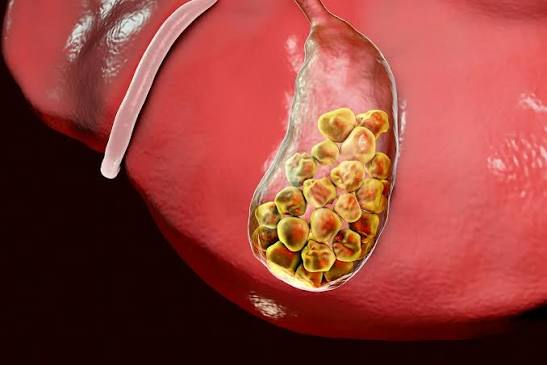
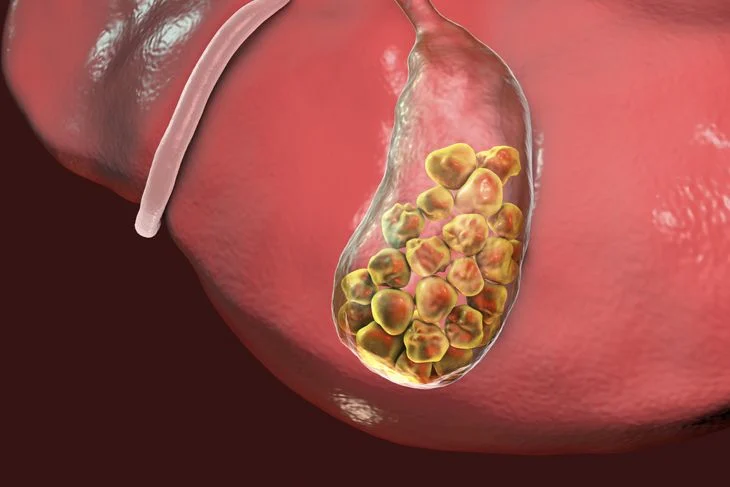
The gallbladder is a small, pear-shaped reservoir tucked beneath the liver. It concentrates the bile the liver produces and releases it into the intestine when a meal containing fat arrives. It is a storage organ, not a manufacturing one — which is why life continues comfortably after it is removed.
Problems begin when bile falls out of balance and forms stones, or when the outlet becomes blocked. A stone that lodges in the cystic duct causes biliary colic: a hard, gripping pain under the right ribs that builds over an hour and often follows a rich meal. If the blockage persists, the wall becomes inflamed and infected, and the picture turns from painful to urgent.
Not every gallstone needs an operation. Stones found by chance, causing no symptoms, are usually watched. Stones that have caused pain tend to cause it again, and in that setting removal of the gallbladder is the definitive answer — done laparoscopically, through four small incisions, with most patients home the same day.
Discuss Your ReportsNone of these confirms a diagnosis on its own. Each is a reason to be examined rather than to wait and see how things develop.
Severe, gripping pain in the upper right abdomen that builds over an hour and often radiates to the right shoulder blade, classically after a fatty meal.
Sickness accompanying the pain, sometimes with retching that brings no relief until the stone shifts back out of the duct.
A rising temperature with the pain suggests the gallbladder wall is inflamed or infected, and needs same-day assessment rather than a wait-and-see approach.
Yellowing of the eyes or skin with dark urine and pale stool means a stone has moved into the bile duct and is obstructing bile flow.
Bloating, belching and discomfort after oily or fried food, often dismissed as ordinary indigestion for months before the diagnosis is made.
A dull, recurring ache below the ribs between attacks, typically worse in the evening and easy to attribute to something else.
Understanding what drives the condition is what makes prevention and early detection practical rather than theoretical.
Only the investigations that will change a decision are ordered, and they are sequenced so results build on one another rather than repeating.
Liver function tests, bilirubin and inflammatory markers separate simple colic from cholecystitis or duct obstruction.
The first-line study. It shows stones, wall thickening and duct dilatation with high accuracy and no radiation.
Reserved for complications — perforation, abscess or an unclear acute abdomen — rather than for finding stones.
Non-invasive magnetic resonance imaging of the bile ducts, used when a stone is suspected to have passed into the common bile duct.
A functional study measuring how well the gallbladder empties, used when pain is typical but ultrasound shows no stones.
Treatment is matched to the stage and to the individual. Not every option below applies to every patient, and the right choice is made together.
Analgesia, antibiotics where infection is present, and dietary modification stabilise an acute attack and buy time for planned surgery.
When a stone has migrated into the common bile duct, endoscopic retrograde cholangiopancreatography clears it and relieves the obstruction before surgery.
The definitive treatment. Four keyhole incisions, a critical-view dissection to protect the bile duct, and same-day discharge for most patients.
Chosen deliberately where dense adhesions, severe inflammation or unclear anatomy make keyhole dissection unsafe. Conversion is a judgement, not a failure.
Specialist surgical care, delivered close to home, without asking families to relocate for months of treatment.
A dedicated hepato-pancreato-biliary and gastrointestinal surgical team, operating on these conditions as their principal practice rather than occasionally.
Endoscopy, endoscopic ultrasound, MRCP and protocol-specific cross-sectional imaging interpreted by clinicians who treat the disease themselves.
Fully equipped theatres, high-dependency support and endoscopy suites, delivered in partnership with hospitals close to where patients live.
Laparoscopic and endoscopic approaches wherever they are the right choice, with shorter stays, less pain and a faster return to ordinary life.
Reports reviewed before you travel, decisions explained in your own language, and a plan built around your circumstances rather than a protocol.
Acute pancreatitis, cholangitis and obstructive jaundice do not keep office hours. Our team is reachable when they present.

Dr. Kapoor leads Advitya's gastrointestinal and hepato-pancreato-biliary practice, with a focus on complex pancreatic, biliary and liver surgery. Her work centres on bringing specialist-level surgical care to regional hospitals, so that patients can receive the operation they need without leaving their families behind.
Pancreato-Biliary-Liver Surgery · Gastrointestinal Surgery · Surgical Oncology
Four steps, explained before they happen, so you always know what comes next.
We begin with your history and your existing reports. Bring every scan and biopsy — they shape the plan more than the symptoms do.
Only the investigations that will change a decision are ordered, sequenced so that you are not sent back and forth between departments.
Complex cases are discussed jointly by surgery, oncology and radiology, and the plan is explained to you and your family in full before anything begins.
Structured recovery with nutritional and physiotherapy support, and a defined follow-up schedule so nothing is left to chance.
Straight answers to the questions patients ask most often in clinic.
The classic symptom is biliary colic — severe pain in the upper right abdomen that builds over an hour, often after a fatty meal, and may radiate to the right shoulder blade. Nausea, vomiting, bloating and fatty food intolerance are common. Fever, chills or yellowing of the eyes indicate a complication and need urgent assessment.
Ultrasound is the first and usually the decisive test — it detects gallstones, wall thickening and duct dilatation accurately and without radiation. Blood tests assess liver function and infection. If a stone may have passed into the bile duct, MRCP provides a detailed non-invasive view before any intervention is planned.
No. Stones discovered incidentally that have never caused symptoms are generally observed rather than removed. Once stones have caused pain, they tend to recur, and cholecystectomy is recommended. Large stones, a porcelain gallbladder or polyps over one centimetre are also grounds for surgery even without symptoms.
It is one of the most commonly performed operations in the world and is very safe in experienced hands. The key safeguard is a careful critical-view dissection to identify the anatomy before anything is divided, which is how bile duct injury is avoided. Conversion to open surgery is occasionally the safest choice and is planned for, not feared.
Most patients go home the same day or the next morning. Desk work is usually resumed within a week, and normal activity including lifting within two to three weeks. Recovery after emergency surgery for acute cholecystitis takes longer, typically three to four weeks.
Yes. The liver continues producing bile, which then drains continuously into the intestine instead of being stored. Most people notice no difference. A minority find very fatty meals loosen the bowel in the first few months, and this settles with modest dietary adjustment.
Connect with our experienced specialists for personalised diagnosis and treatment.
+91 9211221551 · info@advityahealthcares.com · Reports reviewed before you travel